Back Muscles Diagram - Muscle Anatomy Workout Image - weighteasyloss.com - Sore or tenderness in your lower back.. Pain that radiates to your legs, buttock, or thigh areas. Superficial, intermediate, deep and deepest layers.these muscles lie on each side of the vertebral column, deep to the thoracolumbar fascia they span the entire length of the vertebral column, extending from the cranium to the pelvis These muscles include the large paired muscles in the lower back, called erector spinae, which help hold up the spine, and gluteal muscles. Lower back muscle diagram anatomy does degenerative disc disease affect the lower back muscle? Three types of back muscles that help the spine function are extensors, flexors and obliques.
Below you'll see diagrams along with the names of the back muscles that may be the cause of your pain. Others, like sumo deadlifts, have been shown in emg studies—and in the trenches—to focus more on other muscle groups than the back. Both the deltoid and the trapezius are firmly attached to the spine of the scapula. See back muscles and low back pain. We think this is the most useful anatomy picture that you need.
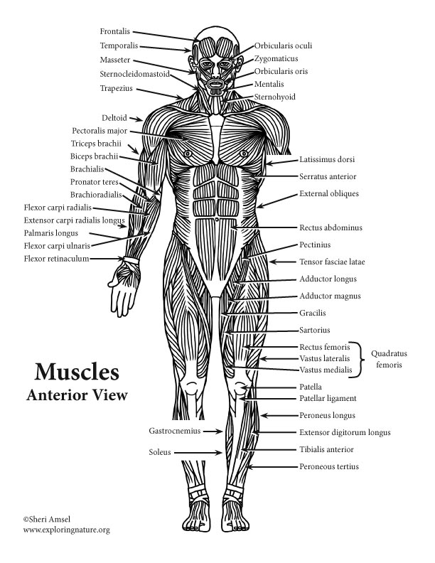
Shoulder Muscles Diagram Labeled - 25 best muscle_blank ... from www.exploringnature.org The trapezius and latissimus dorsi muscles connect the upper limb to the vertebral column. Symptoms of muscle pain include: Back muscles, back muscle diagram. The most common symptoms of a torn muscle, strained muscle, and pulled muscle include: For more anatomy content please follow us and visit our website: In this image, you will find 1st cervical vertebrae, atlus, cervical plexus, 7th cervical vertebrae, 1st thoracic vertebrae, brachial plexus, spinal dura mater, filaments of spinal nerve roots, 12th thoracic vertebra, 1st lumber vertebra, iliohypogastric nerve, ilioinguinal nerve, lumbar. See back muscles and low back pain. What is the origin and insertion of the rhomboid minor and major muscle?
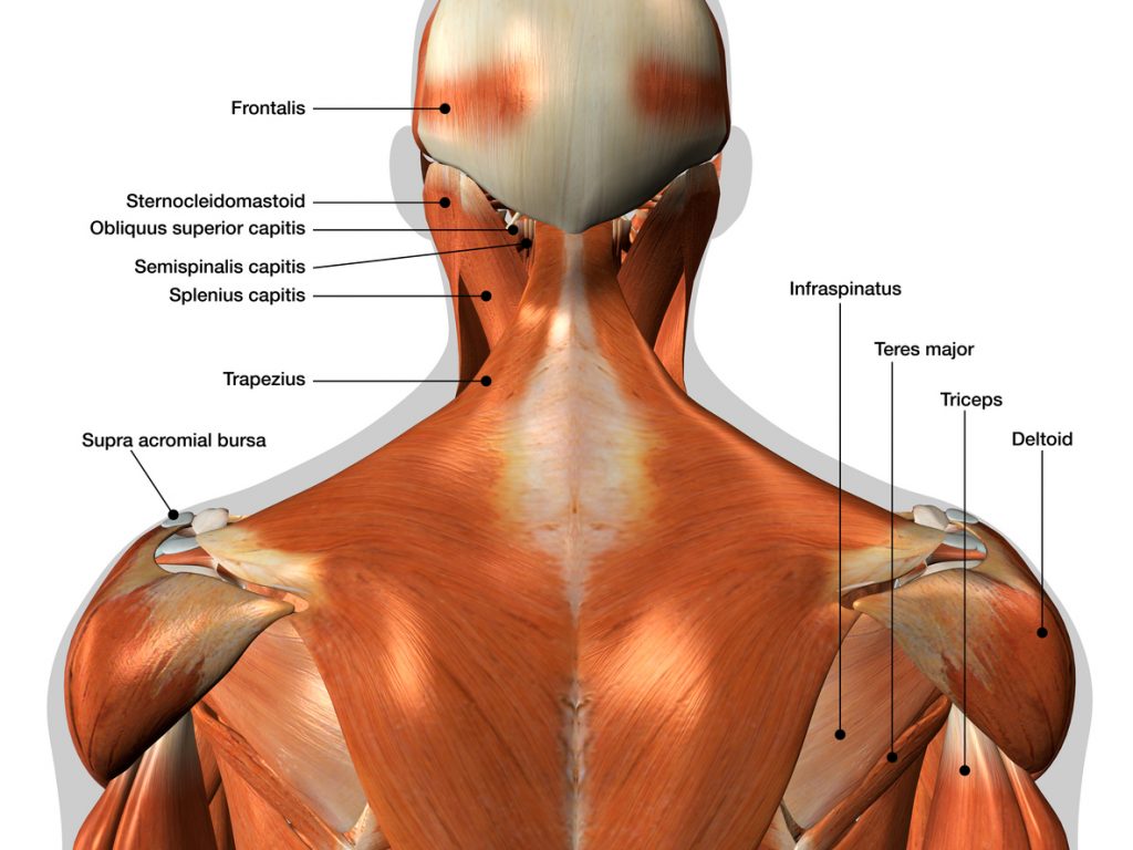
The deltoid, teres major, teres minor, infraspinatus, supraspinatus (not shown) and subscapularis muscles (not shown) all extend from the scapula to the humerus and act on the shoulder joint.
Muscles of the lower back and buttocks diagram, human muscles, muscles of the lower back and buttocks diagram. Muscle spasms (contraction or stiffening of the back muscles) muscles that feel tight; What is the origin and insertion of the rhomboid minor and major muscle? Working the lower back, erector spinae muscles, and hamstrings, a barbell deadlift requires back strength to effectively complete. The part of the nerve that emerges out of the spine is called the nerve root. Superficial back muscles, intermediate back muscles and intrinsic back muscles.the intrinsic muscles are named as such because their embryological development begins in the back, oppose to the superficial and intermediate back muscles which develop elsewhere and are therefore classed as extrinsic muscles. The back consists of the spine, spinal cord, muscles, ligaments, and nerves. Back muscles chart, back muscles diagram and ligaments, back muscles diagram lats, back muscles diagram massage, upper back muscles chart, human muscles, back muscles. We hope this picture anatomy of back muscles diagram can help you study and research. Another common cause of lower back and hip pain is disc injury. The muscles of the back are a group of strong, paired muscles that lie on the posterior aspect of the trunk they provide movements of the spine, stability to the trunk, as well as the coordination between the movements of the limbs and the back muscles are divided into two large groups: This is a diagram of the larger and more surface muscles of the low back. Sore or tenderness in your lower back.
The extrinsic (superficial) back muscles, which lie most superficially on the back. The pelvis at the bottom of the back and the shoulders at the top of the back give the back. The trapezius and latissimus dorsi muscles connect the upper limb to the vertebral column. Daniel nelson on january 1, 2019 2 comments 🔥! We hope this picture anatomy of back muscles diagram can help you study and research.
Anatomy of the intrinsic back muscles. Line diagram of ... from www.researchgate.net Superficial back muscles, intermediate back muscles and intrinsic back muscles.the intrinsic muscles are named as such because their embryological development begins in the back, oppose to the superficial and intermediate back muscles which develop elsewhere and are therefore classed as extrinsic muscles. The back has a total of 40 muscles. Most of the time, back muscle pain is diagnosed then treated with little more than a prescription of rest, painkillers and muscle relaxants. The pelvis at the bottom of the back and the shoulders at the top of the back give the back. Creatine is now proving to be one of the most potent muscle growth accelerators giving excellent muscle mass increase and phenomenal strength increases order yours today. To learn more about the anatomy of the spine, watch this video. The human back extends from the buttocks to the posterior portion of the neck and shoulders. Lower back muscle diagram anatomy does degenerative disc disease affect the lower back muscle?
Three types of back muscles that help the spine function are extensors, flexors and obliques.
For more anatomy content please follow us and visit our website: To learn more about the anatomy of the spine, watch this video. Superficial back muscles, intermediate back muscles and intrinsic back muscles.the intrinsic muscles are named as such because their embryological development begins in the back, oppose to the superficial and intermediate back muscles which develop elsewhere and are therefore classed as extrinsic muscles. How many muscles are in the back? Back muscles, back muscle diagram. The most common symptoms of a torn muscle, strained muscle, and pulled muscle include: When back development is the goal, stick to one of these variations. Lower back muscle diagram anatomy does degenerative disc disease affect the lower back muscle? See back muscle anatomy stock video clips. Muscles of the back diagram. Muscle strain is often the cause of back pain from heavy lifting or vigorous exercise. And reach, pull and extend your arms and torso. Stand behind the barbell with your feet shoulder.
The human back extends from the buttocks to the posterior portion of the neck and shoulders. See back muscles and low back pain. Daniel nelson on january 1, 2019 2 comments 🔥! The back consists of the spine, spinal cord, muscles, ligaments, and nerves. Superficial, intermediate, deep and deepest layers.these muscles lie on each side of the vertebral column, deep to the thoracolumbar fascia they span the entire length of the vertebral column, extending from the cranium to the pelvis
Back Muscles Diagram : Image result for back muscles ... from www.ericfavre.com Pain that radiates to your legs, buttock, or thigh areas. To learn more about the anatomy of the spine, watch this video. The back consists of the spine, spinal cord, muscles, ligaments, and nerves. Back muscles chart, back muscles diagram and ligaments, back muscles diagram lats, back muscles diagram massage, upper back muscles chart, human muscles, back muscles. Stiffness in the back region. Three types of back muscles that help the spine function are extensors, flexors and obliques. What is the origin and insertion of the rhomboid minor and major muscle? Working the lower back, erector spinae muscles, and hamstrings, a barbell deadlift requires back strength to effectively complete.
Support and protect your spine;
Working the lower back, erector spinae muscles, and hamstrings, a barbell deadlift requires back strength to effectively complete. Anatomynote.com found anatomy of back muscles diagram from plenty of anatomical pictures on the internet. The back has a total of 40 muscles. Likewise, there are muscles in other parts of the body that help support and move the spine. Below you'll see diagrams along with the names of the back muscles that may be the cause of your pain. Learn vocabulary, terms, and more with flashcards, games, and other study tools. Five pairs of lumbar spinal nerves labeled l1 to l5 branch off your spinal cord and exit through small holes between the vertebrae. This muscle is a major generator of lower back and hip pain, as well as being responsible for complaints of a burning sensation along the posterior superior iliac spine (psis) and sacroiliac joint. The extensor muscles are attached to back of the spine and enable standing and lifting objects. The trapezius and latissimus dorsi muscles connect the upper limb to the vertebral column. It is opposite from the chest, and the vertebral column runs down the back. In this image, you will find 1st cervical vertebrae, atlus, cervical plexus, 7th cervical vertebrae, 1st thoracic vertebrae, brachial plexus, spinal dura mater, filaments of spinal nerve roots, 12th thoracic vertebra, 1st lumber vertebra, iliohypogastric nerve, ilioinguinal nerve, lumbar. The muscles of the back are a group of strong, paired muscles that lie on the posterior aspect of the trunk they provide movements of the spine, stability to the trunk, as well as the coordination between the movements of the limbs and the back muscles are divided into two large groups: